Development Year: 1971
Inventor: Raymond Vahan Damadian
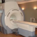
Description: Magnetic resonance imaging (MRI) is a medical imaging technique used in radiology to form pictures of the anatomy and the physiological processes of the body. MRI scanners use strong magnetic fields, magnetic field gradients, and radio waves to generate images of the organs in the body.
The MRI is widely used in hospitals and clinics for medical diagnosis, staging and follow-up of disease diagnosis due to its ability to provide sharp contrast in images of soft tissues like the brain and abdomen.
Functionality: Strong magnetic field created by the MRI scanner causes the atoms in your body to align in the same direction. Radio waves are then sent from the MRI machine and move these atoms out of the original position. As the radio waves are turned off, the atoms return to their original position and send back radio signals. These signals are received by a computer and converted into an image of the part of the body being examined.
Source: https://www.mayoclinic.org/tests-procedures/mri/about/pac-20384768#:~:text=Magnetic%20resonance%20imaging%20(MRI)%20is,large%2C%20tube%2Dshaped%20magnets.